Label the anatomical features of the forearm and wrist – Labeling the anatomical features of the forearm and wrist is a crucial step in understanding the intricate workings of the human musculoskeletal system. This comprehensive guide delves into the intricacies of the forearm and wrist, providing a detailed exploration of their bones, muscles, arteries, veins, nerves, and surface anatomy.
By delving into the depths of this topic, we uncover a wealth of knowledge that is essential for students, medical professionals, and anyone seeking a deeper understanding of human anatomy.
1. Radius and Ulna Bones

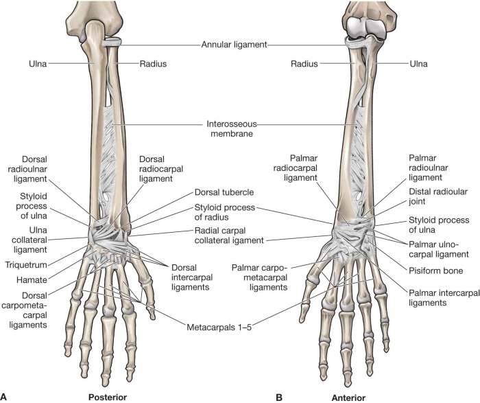
The radius and ulna are two long bones that form the forearm. The radius is located on the lateral side of the forearm, while the ulna is located on the medial side.
The radius and ulna articulate with each other at the proximal and distal ends of the forearm. At the proximal end, the radius articulates with the capitulum of the humerus, while the ulna articulates with the trochlea of the humerus.
At the distal end, the radius articulates with the carpal bones, while the ulna articulates with the triangular fibrocartilage complex.
The radius and ulna allow for a wide range of movements at the forearm, including flexion, extension, pronation, and supination.
Anatomical Landmarks, Label the anatomical features of the forearm and wrist
- Radial head: The proximal end of the radius that articulates with the capitulum of the humerus.
- Olecranon process: The proximal end of the ulna that articulates with the trochlea of the humerus.
- Trochlear notch: A notch on the anterior surface of the ulna that articulates with the trochlea of the humerus.
2. Wrist Bones (Carpals): Label The Anatomical Features Of The Forearm And Wrist

The wrist is made up of eight carpal bones that are arranged in two rows.
The proximal row of carpal bones consists of the scaphoid, lunate, triquetrum, and pisiform bones. The distal row of carpal bones consists of the trapezium, trapezoid, capitate, and hamate bones.
The carpal bones form the carpal tunnel, which is a narrow passageway through which the median nerve and flexor tendons pass.
Shape, Location, and Function of Each Carpal Bone
- Scaphoid: A boat-shaped bone located on the radial side of the proximal row.
- Lunate: A crescent-shaped bone located on the radial side of the proximal row.
- Triquetrum: A triangular-shaped bone located on the ulnar side of the proximal row.
- Pisiform: A small, pea-shaped bone located on the anterior surface of the triquetrum.
- Trapezium: A trapezoidal-shaped bone located on the radial side of the distal row.
- Trapezoid: A trapezoidal-shaped bone located on the radial side of the distal row.
- Capitate: A large, central bone located in the distal row.
- Hamate: A hook-shaped bone located on the ulnar side of the distal row.
FAQ Explained
What are the major bones of the forearm?
The major bones of the forearm are the radius and ulna.
How many carpal bones are there?
There are eight carpal bones arranged in two rows.
What is the function of the forearm muscles?
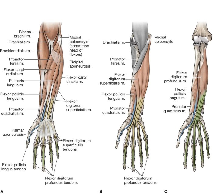
The forearm muscles contribute to flexion, extension, pronation, and supination of the forearm.
Which nerves innervate the forearm?
The major nerves of the forearm include the median, ulnar, and radial nerves.

